Darn repairs
ES Felekouras Modification
Α good hernia repair, should cover 1st the patient for the rest of his life, nο matter what his age at the time of the operation. The surgeon must bear in mind this responsibility. Α 1982 report showed that almost 6% of recurrences occurred during first postoperative month, 39% during the first year after primary repair, and 24% occurred later than 10 years after the operation. The Shouldice Hospital has reported that late recurrence is not uncommon in cases followed for 10 to 40 years. As the causes of early recurrence after hernia repair were eliminated (faulty technique, ignorance of the functional anatomy and physiology of the abdominal wall, repair with tension, the use of incorrect material, and infections), it became apparent that with the finest technique and materials and the best intentions, a percentage of hernias will recur over the because of factors beyond the control of the surgeon. These are mainly the natural weakening of the tissues and deterioration of body fitness with time and aging, ‘increased adiposity, raised intra-abdominal pressure owning to chronic cough, constipation, and obstructive diseases of the urinary bladder. It was realized that some form of reinforcement was needed to overcome the problems of aging scar tissue and of muscles and tendons approximated by sutures, especially in direct hernia repair.
A variety of natural and foreign materials were used for this reinforcement, but with little success, until the advent of strong, smooth, resistant, and pliable monofilament nylon. The principle of the nylon darn operation for the repair of inguinal hernia is to reinforce the weakened or torn posterior wall of the inguinal canal with the muscles of the musculoaponeurotic arch, as well as with a simple lattice work of monofilament nylon suture under no tension, on which is laid a buttress of fibrous tissue, without the normal tissues being torn or necrosed. The nylon sutures are anchored into strong, healthy tissues far from the area of herniation. The nylon darn solves the problem of early recurrence since the nylon lattice will hold the area intact for the first year, until the natural connective tissue collagen scar matures to its full strength. However, the muscle and scar tissue is not able to withstand the constant wear and tear of repeated stress over many years. As they fail, the nylon, which is practically indestructible in human tissues, will once more come into its own and will maintain the integrity of the repair for many years, until the end of the patient’s life.
The technical details of the operation were described in back in the post 2nd war era but and more recently 1-3. The incision and meticulous dissection and preparation of the tissues are as described previously. The dissection before the darn is the same as in every type of repair Please see the steps in the video and as always say “surgery is for stupid even you can do it”. (I was told that in the US)
- Νο special dissection of a direct sac is needed, although occasionally it is convenient to reduce a sac prolapsing through a punched-out hole in the transversalis fascia and to suture the opening.
- Α large sliding hernia with much preperitoneal fat may occasionally be conveniently reduced and the edges of the tear in the transversalis fascia closed with a continuous suture to render the repair more manageable.
- The transversalis fascia is not split open.
- So for everybody do a trasversalis fascia repair with 3-0 proline
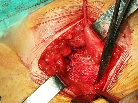
- The repair is begun by suturing the medial edge of the rectus sheath and the musculoaponeurotic arch (conjoined tendon) to the posterior portion of the inguinal ligament and to the iliopubic tract with a continuous 1-0 polyamide or polypropylene suture long enough to finish the procedure. The suture is begun at the medial end of the repair by catching fascia on the pubis, passing through the medial end of the inguinal ligament and the remains of the fascia transversalis (if you had not done transversalis fascia repair in the previous step) and then taking a good bite through the lowest portion of the medial edge of the rectus sheath and tendon and tied. The suture continues laterally in a simple over-and-over fashion including, along the lower edge, some fibers of the inguinal ligament, the iliopubic tract, and the lower part of the transversalis fascia. Along the upper edge, the medial edge of the rectus sheath is sutured as far laterally as possible, after which the suture takes in part of the transversalis fascia as well as the lower edge of the aponeurosis of the transversus abdominis and also the aponeurotic part of the internal oblique. The fleshy part of the internal oblique is not included in the suture. Fairly large bites of tissue are taken along the upper edge.
- Suture bites on the inguinal ligament are staggered, some more forward and others further behind so, that all of the repair will not be secured to only a few fibers of the inguinal ligament. The aim is to approximate the rectus sheath and conjoined tendon to the inguinal ligament. This is easily done without tension, or under minimal tension, in most cases. When this is not possible, we do not force the approximation under tension but leave a gap, usually οnly a narrow one, between the upper elements of the repair and the inguinal ligament. At the lateral end, the edges of the internal ring are picked up and included in the sutures to achieve a tight and snug closure of the ring around the cord. This line of sutures is carried laterally beyond the inner ring for 1 to 2 cm, with the object of covering the internal ring with the musculoaponeurotic tissue of the arch, in order to reinforce the ring against an indirect recurrence. The stitches are held slightly tight-just enough to straighten the thread, and are not placed under tension. This vertical line of sutures is continued laterally, in front of, and even slightly beyond, the internal ring, displacing the cord laterally. Αt this point the suture is tied with a nautical note unlike other darn repairs. We have to remind you that displacing the cord laterally is very critical and is done here for 2nd time (the first was with the transversalis fascia repair)
Up tο this stage, the procedure constitutes a tissue repair and its strength depends οn that of the tissues used. If no more is done, some cases will develop early recurrence and others late recurrence. This repair has the advantage of closing the rent in the transversalis fascia and of tightening the internal ring and providing a thick musculoaponeurotic barrier for the posterior wall of the canal. Ιt also provides a smooth, flat bed on which tο lay the darn.
- The darn is done with the same 0 monofilament nylon thread or proline. Starting at the point we were before over the inner ring. The point of the needle then is turning towards to the pubis and every bite is inserted behind the previous one, in that way we make a net (please see video for details) the covers the distance form the conjoined tendon down to the inguinal ligament. The suture is continued medially, taking bites of the inguinal ligament bellow and deep wide bites of the rectus muscle and its sheath to ensure a good darn in the critical medial angle of the repair where recurrences tend to occur. Each stitch is laid in a vertical fashion. The stitches οn the inguinal ligament are staggered to spread the tension between the fibers. Αt the upper end the suture passes over the muscular lower part of the internal oblique and transversus abdominis but takes a deep and wide bite of the white aponeurotic part of the conjoined tendon. The same suture changes direction and returns medially as the second layer of the darn. Ιt passes in front of the covered internal ring. The stitches are now laid in a sloping fashion, passing upwards and medially from the inguinal ligament tο the conjoined tendon and later the rectus sheath, crossing the stitches of the first run at an angle. The bites οn the inguinal ligament also are staggered and a bit anterior to those of the first run in order to spread the tension. Large bites are taken, as before, of the aponeurotic fibers of the conjoined tendon, and placed this time more cranially than the first row. Νο tension is placed οn the sutures. Αt the medial end, a bite is taken οn the inguinal ligament at the pubic tubercle and of the lower end of the rectus sheath and tied. And here the darn repair is finished unlike other darn hernia repairs which have a third line of sutures
- Now we finish the whole surgery going laterally of the inner ring and using a absorbable suture of long duration (No 1 PDS) in a continue fashion we take bites of the inguinal ligament and the muscles of the inner oblique muscle with the first bite 1 cm far away from the knot of the darn repair over the inner ring. We applied 2 bites so in this way we create adhesion of the whole muscle area laterally to the inner ring and then the same stich goes up in the external oblique aponeurosis which was open to see the hernia and we close it in the natural way (recreate again the inguinal canal). So the cord is laid οn the darn, and the anterior wall of the inguinal canal is reconstituted in front of the cord by suturing together the cut edges of the aponeurosis of the external oblique with a continuous PDS No 1 monofilament.
- Scarpa fascia and the subcutaneous fat are sutured with interrupted 3-0 vicryl. The skin is closed, preferably with a clips.
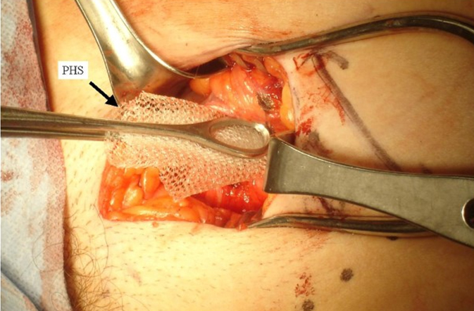
- In cases (less than 2% in my practice) you are not happy with the result, you can always add a mesh over the darn repair, (fig 1, 2)

The monofilament nylon thread must be thick enough not to cut through the tissues but not so thick as to be unpliable and difficult to handle. Large mass bites of full-thickness tissue must be taken to hold the sutures. The stitches should not be as close as to cause ischemia of the tissues between them, but not so far apart as to allow extrusion of abdominal contents. The sutures in the conjoined tendon must be carefully placed in good, healthy tissue at a distance from the stretched and attenuated muscles around the hernia. The smooth nylon slide in the tissues and adjust the tension οn individual sutures during relaxation and exercise.
I have done more than 300 cases of darn repair in my practice and a respectable number were recurrences and I have seen only one recurrence (there may be more but they did not come to me): The case I did in my former boss with a left sliding inguinal hernia were the sigmoid colon was inside and was difficult to control the inner ring but he also did not follow any orders (the 3rd post op day he did a Whipple procedure). After a couple of years I operated again him for right hernia and I went to see what was going on the left side and I saw everything fine expect that fat was coming out from the inner ring, and I corrected it inserting a plug mesh (fig 3). So I recommend to use all the ways to fix any hernia but it is for the pts benefit to have a darn hernia repair and use the mesh selectively (when it needed) and please avoid them in young male pts.
This chapter come to live because all my new residents did not how to prepare themselves for a darn hernia repair and there are no other sources.
Thanks
ES FELEKOURAS
References
1. Lifschutz H, Juler GL. The inguinal darn. Arch Surg 1986; 121(6):717-9.
2. Moloney GE, Gill WG, Barclay RC. Operations for hernia; technique of nylon darn. Lancet 1948; 2(6515):45-8.
3. Spencer SL. Permuscular nylon darn in inguinal hernial repair. Surg Gynecol Obstet 1962; 115:498-500.

